MLZ is a cooperation between:
MLZ is a member of:
 > ERF-AISBL
MLZ on social media:


MLZ (eng)
Lichtenbergstr.1
85748 Garching
ANTARES
Cold neutron radiography and tomography facility

This instrument is focussed on cold neutrons. Therefore, please carefully check the “Technical data WITHOUT cold source” section. Deviating parameters are in bold. The instrument team is happy to answer any further questions!
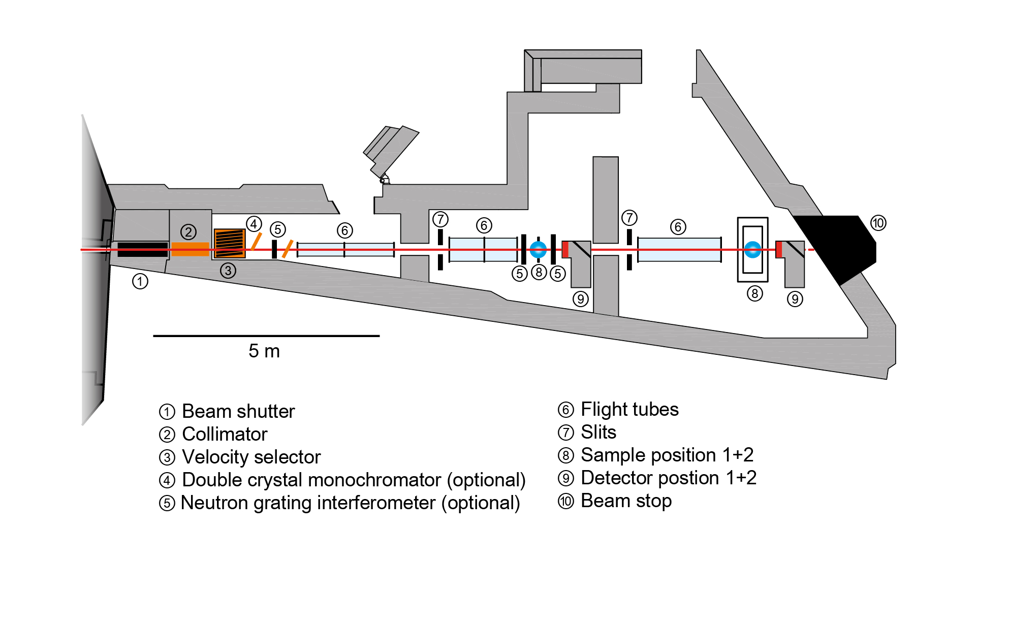
The neutron imaging facility ANTARES is located at the neutron beam port SR-4a. Based on a pinhole and flight tube principle (no neutron guide) with a variable collimator located close to the beam port, the facility provides for flexible use in high-resolution and high-flux imaging.
ANTARES offers two different detector positions, which may be chosen according to the requirements for beam size, neutron flux and spatial resolution. Both chambers offer abundant space for user-provided experimental systems or sample environments.
The beam formation chamber is separately accessible for the optional installation of devices shaping the beam and spectrum provided by the user. At this position, ANTARES also offers built-in options such as a velocity selector, a neutron grating interferometer and a double crystal monochromator, which are readily available for standard user operation.
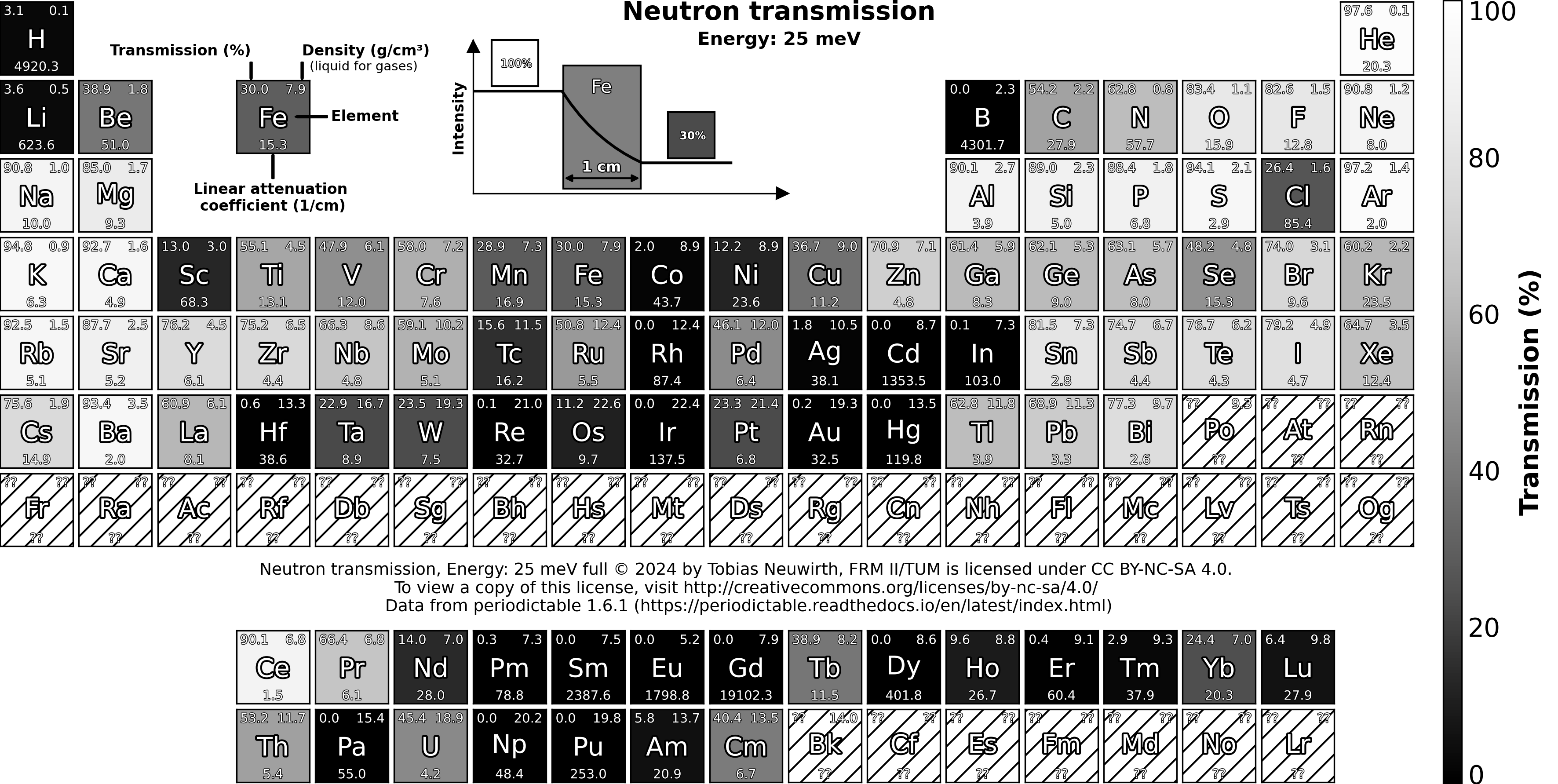
The spectrum of ANTARES is shown in fig. 1 (see gallery). ANTARES offers a high neutron flux of approx. 6.3 × 10⁷ n cm-2 s-1 at a typical collimation of L/D = 500. By using different pinholes, the collimation can be adjusted to the experiment’s requirements. Additionally, we can provide access to a microfocus X-ray CT set-up for complementary investigations with a spatial resolution as good as 1 μm.
The neutron imaging facility ANTARES is designed to deliver radiographs and CTs of samples, often complementary to X-ray measurements. The most important features are the high penetration of metals (Fe ~ 4 – 5 cm, Al ~ 20 – 30 cm, Pb ~ 10 – 20 cm) and the high sensitivity for hydrogen. These allow to visualise metal machine parts as well as liquids, sealants, and plastics inside of metal parts. Liquid contrast agents can be employed for crack and void detection.
Typical applications are in archaeology, paleontology, biology, life sciences, magnetism, and non-destructive testing of mission-critical parts.
Available techniques:- Neutron radiography of static samples and dynamic processes with up to several 100 frames per second (depending on spatial resolution)
- Computed tomography (a few seconds per scan up to several hours depending on spatial resolution)
- Neutron Grating Interferometry (spatially resolved USANS off structures of 100 nm – 10 µm)
- Bragg Edge Imaging
- Polarised neutron imaging
- Tensile rig: 100 kN
- 3He cryostat: min. 500 mK
- Normal conducting magnet: 300 mT
- Vacuum furnace: up to 400°C
Without the cold source, ANTARES will have a similar flux as before, but the spectrum will be fully thermalised instead of cold. Applications using cold neutrons will be possible up to a wavelength of approx. 4 Å with reduced flux. All other instrument properties will remain unchanged.
Beam properties- Default collimation ratio L/D=500 at the sample position with a flux of 5.4 × 10⁷ n cm-2 s-1
- Collimation ratio adjustable between L/D = 250 and 7200
- Beam size up to 35 × 35 cm²
- Neutron velocity selector: 1.6 Å ≤ λ ≤ 3 Å (Δλ/λ = 20 %) or 3.0 Å ≤ λ ≤ approx. 4 Å (Δλ/λ = 10 %)
- Double crystal monochromator: 1.4 Å ≤ λ ≤ approx. 4.0 Å (1 % < Δλ/λ < 3 %)
- Neutron grating interferometer: Sensitive to USANS scattering on length scales of 100 nm – 10 µm, optimised for thermal neutrons
- Filter for epithermal neutron imaging for higher penetration
- Neutron polarisation analysis set-up using 3He polarisers and thermal neutrons
- Flexible options depending on sample size and weight (up to 500 kg)
- Camera and scintillator-based detection systems with spatial resolutions down to a few µm.
- Standard detector: ANDOR cooled CCD camera, 2048 × 2048 pixels, 16 bit
- Fast, cooled scientific CMOS camera: ANDOR Neo 2560 × 2160 pixels, 16 bit, up to 50 fps full frame
- Intensified high speed CMOS camera, 1920 × 1080 pixels with 2000 fps
- Imaging plate readers with neutron and X-ray imaging plates (focus size 12.5 µm)
Thermal neutron spectrum: See fig. 1
- Default collimation ratio L/D=500 at the sample position with a flux of 6.3 × 10⁷ n cm-2 s-1
- Collimation ratio adjustable between L/D = 250 and 7200
- Beam size up to 35 × 35 cm²
- Neutron velocity selector: 1.6 Å ≤ λ ≤ 3 Å (Δλ/λ = 20 %) or 3.0 Å ≤ λ ≤ 6.5 Å (Δλ/λ = 10 %)
- Double crystal monochromator: 1.4 Å ≤ λ ≤ 6.5 Å (1 % < Δλ/λ < 3 %)
- Neutron grating interferometer: Sensitive to USANS scattering on length scales of 100 nm – 10 µm
- Filter for epithermal neutron imaging for higher penetration
- Neutron polarisation analysis setup using V-cavities or 3He polarisers
- Flexible options depending on sample size and weight (up to 500 kg)
- Camera and scintillator-based detection systems with spatial resolutions down to a few µm.
- Standard detector: ANDOR cooled CCD camera, 2048 × 2048 pixels, 16 bit
- Fast, cooled scientific CMOS camera: ANDOR Neo 2560 × 2160 pixels, 16 bit, up to 50 fps full frame
- Intensified high speed CMOS camera, 1920 × 1080 pixels with 2000 fps
- Imaging plate readers with neutron and X-ray imaging plates (focus size 12.5 µm)
Cold neutron spectrum: See fig. 1
Instrument scientists
Dr. Robin Woracek
Phone: +49 (0)89 289-11692
E-mail: robin.woracek@frm2.tum.de
Dr. Burkhard Schillinger
Phone: +49 (0)89 289-12185
E-mail: burkhard.schillinger@frm2.tum.de
Dr. Tobias Neuwirth
Phone: +49 (0)89 289-11754
E-mail: tobias.neuwirth@frm2.tum.de
ANTARES
Phone: +49 (0)89 289-14815
Operated by
Funding
News

Publications
Find the latest publications regarding ANTARES in our publication database iMPULSE:
Citation templates for users
In all publications based on experiments on this instrument, you must provide some acknowledgements. To make your work easier, we have prepared all the necessary templates for you on this page.
Instrument control
Gallery


MLZ is a cooperation between:
MLZ is a member of:
> ERF-AISBL
MLZ on social media: