MLZ is a cooperation between:
MLZ is a member of:
 > ERF-AISBL
MLZ on social media:


MLZ (eng)
Lichtenbergstr.1
85748 Garching
MARIA
Magnetic reflectometer with high incident angle

This instrument is focussed on cold neutrons. Therefore, please carefully check the “Technical data WITHOUT cold source” section. Deviating parameters are in bold. The instrument team is happy to answer any further questions!
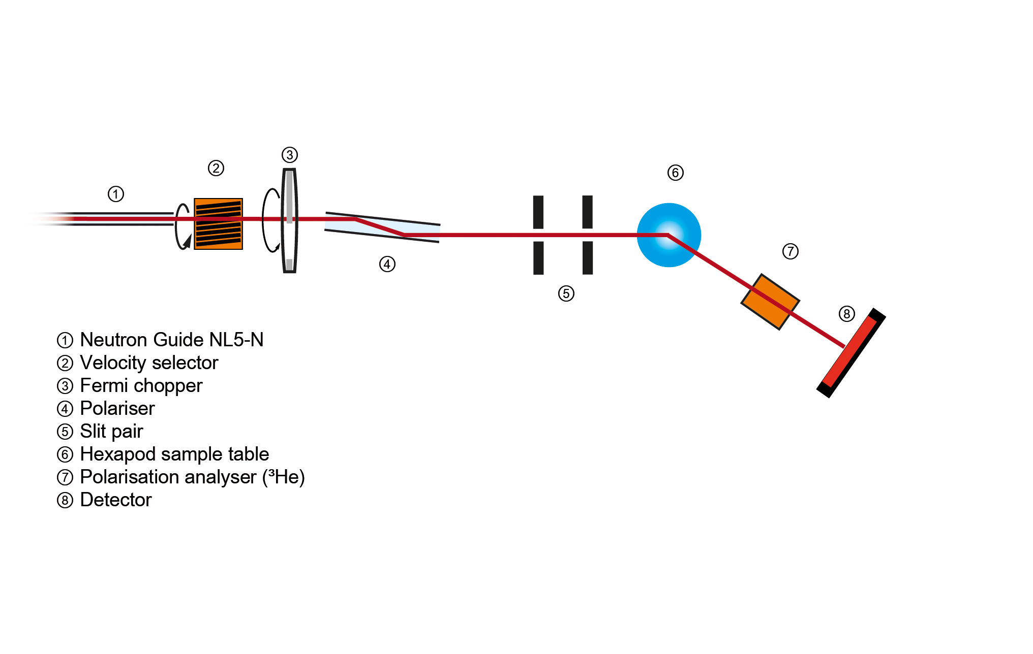
The neutron reflectometer MARIA with polarisation analysis was designed to investigate thin magnetic layered structures down to the monolayer scale and lateral structures. The reflection of polarised neutrons allows us to determine individually the density, the modulus, and the direction of the magnetisation vector of buried layers.
MARIA is optimised for layer thicknesses between 3 – 300 Å and lateral structure sizes from nm to µm. The instrument is working with a small focussed beam and sample sizes of 1 cm2 at λ = 4.5 Å in a vertical orientation with a maximum incident angle of 180° and outgoing angle ranging from -14° to 100°.
MARIA provides polarisation analysis in standard operation, where the beam is polarised by a polarising guide (z-geometry) and analysed by a wide-angle 3He-cell. This can alternatively be done by a supermirror analyser for only the specular beam providing registration of non-spin-flip and spin-flip signal simultaneously.
Besides the above-described reflectometer mode with good resolution in the horizontal scattering plane, MARIA can be used in the GISANS mode with additional resolution in the vertical direction. The latter mode allows for measuring lateral structures down to the nm scale.
With scattering under grazing incidence, we investigate depth-resolved laterally-averaged magnetisations and the correlations between their lateral fluctuations. With an additionally polarised neutron beam, we derive vector information on the laterally-averaged magnetisation (reflectivity) and the correlations between their lateral fluctuations (off-specular scattering – µm length scale, GISANS – nm length scale).
In general, MARIA can be used for measurements of magnetic roughness, the formation of magnetic domains in thin layered structures, lateral structures, etc. (polarised mode) and density profiles, structures of solid polymer layers, etc. (unpolarised mode with higher intensity).
Results possible without the need for multilayer investigation of:- Diluted semiconductors
- Influence of the substrate
- Interfaces between oxide materials
Additionally, the instrument in non-polarised beam mode can be used for reflectometry and GISANS studies of ‘soft’ layers at the solid/ liquid interface by the use of appropriate liquid cells that are available at the beamline. Candidate systems for such investigations include polymer brushes, polyelectrolyte multilayers, biomimetic-supported membranes, adsorbed proteins etc. For typical applications involving deuterated solvents, the dynamic range that can be expected covers six orders of magnitude.
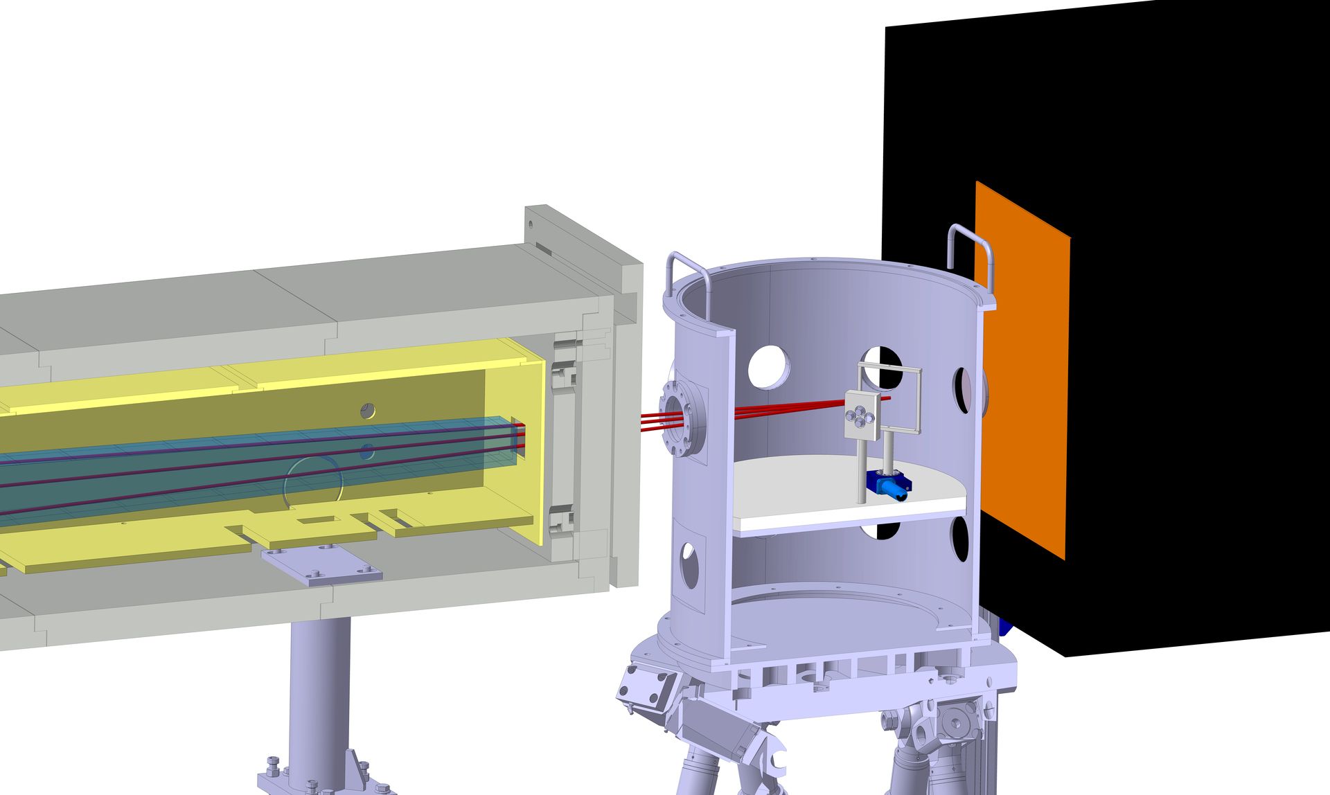
- Hexapod with additional turntable (360°) at sample position: max. load up to 500 kg
- Bruker electromagnet: up to 1.3 T
- 3He closed cycle cryostat: down to 4 K
Besides this standard set-up the complete sample environment of the JCNS can be adopted to MARIA so that magnetic fields up to 5 T and temperatures from 50 mK to 500 K are available.
All parts of MARIA are controlled by a computer system according to the ‘Jülich-Munich’ standard based on a Linux workstation. This allows a flexible remote control with automatic scan programs, including the control of sample environments such as cryostat and electromagnet.
Furthermore, MARIA can provide users with a fully equipped Oxid-MBE. The typical sample sizes are 10 × 10 mm2 and as targets we can provide Al, Cr, Pr, Fe, La, Nb, Ag, Nd, Tb, Sr, Mn, Ti and Co.
- Neutron guide NL5-N:
- Vertically focussing elliptic guide
- Monochromator: Velocity selector
- Wavelength:
- 4.5 Å – 10 Å (polarised)
- 4.5 Å – 40 Å (unpolarised)
- Resolution:
- 10 or 20% velocity selector
- 1 %, 3 % Fermi chopper
- Double reflection polariser
- Horizontal scattering plane
- Expected polarised flux 5 × 10⁶ n cm-2 s-1 for 3 mrad collimation
- Distance S1 – S2 (collimation): 4100 mm
- Distance S2 – sample: 400 mm
- Max. opening S2: 50 mm × 40 mm (w × h)
- Distance sample – detector: 1910 mm
- Max. detector angle: 120°
- GISANS option: 4 m collimation length
- Reflectometry:
- Qz- range: 0.002 Å-1 – 3.2 Å-1
- Qx- range: 6 × 10⁻⁵ Å-1 – 0.001 Å-1
- αf -14° – 100°
- GISANS option:
- Qy- range: 0.002 Å-1 – 0.2 Å-1
- 3He cell (off-specular and GISANS)
- Supermirror analyser (specular reflectivity)
- Neutron guide NL5-N:
- Vertically focussing elliptic guide
- Monochromator: Velocity selector
- Wavelength:
- 4.5 Å – 10 Å (polarised)
- 4.5 Å – 40 Å (unpolarised)
- Resolution:
- 10 or 20% velocity selector
- 1 %, 3 % Fermi chopper
- Double reflection polariser
- Horizontal scattering plane
- Expected polarised flux 5 × 10⁷ n cm-2 s-1 for 3 mrad collimation
- Distance S1 – S2 (collimation): 4100 mm
- Distance S2 – sample: 400 mm
- Max. opening S2: 50 mm × 40 mm (w × h)
- Distance sample – detector: 1910 mm
- Max. detector angle: 120°
- GISANS option: 4 m collimation length
- Reflectometry:
- Qz- range: 0.002 Å-1 – 3.2 Å-1
- Qx- range: 6 × 10⁻⁵ Å-1 – 0.001 Å-1
- αf -14° – 100°
- GISANS option:
- Qy- range: 0.002 Å-1 – 0.2 Å-1
- 3He cell (off-specular and GISANS)
- Supermirror analyser (specular reflectivity)
Neutron Depth Profiling (NDP)
Alternatively to the reflectometer/GISANS mode, MARIA can be operated with a multidetector Neutron Depth Profiling (NDP) set-up using the focussed beam.
NDP allows the quantitative determination of the in-depth distribution for several light elements such as 6Li, 10B, 14N, and a few others. In this instrument operation mode, the NDP chamber is placed on the sample table, placing the NDP sample in the focal point of the vertically focussing elliptic neutron guide. The thermal equivalent neutron flux at the sample position amounts to 0.7 × 10⁸ n cm-2 s-1. Combined with a multiple detector system for charged particle collection, it gives the opportunity to perform fast, time-resolved NDP measurements with a depth resolution of 10 nm.
Different types of solid-state Li-batteries and battery materials.
- Expected thermal equivalent flux: 0.7 × 10⁸ n cm-2 s-1 (10 × 20 mm2)
- Nominal limit of quantisation for 6Li ~ 1013 at/cm2
- Energy resolution (alpha-particles): < 11 keV
- Expected thermal equivalent flux: 7 × 10⁸ n cm-2 s-1 (10 × 20 mm2)
- Nominal limit of quantisation for 6Li ~ 1013 at/cm2
- Energy resolution (alpha-particles): < 11 keV
Instrument scientists
Dr. Alexandros Koutsioumpas
Phone: +49 (0)89 158860-674
E-mail: a.koutsioumpas@fz-juelich.de
Dr. Sabine Pütter
Phone: +49 (0)89 158860-742
E-mail: s.puetter@fz-juelich.de
MARIA
Phone: +49 (0)89 158860-514
Neutron Depth Profiling (NDP)
N/a
Operated by

Funding

Publications
Find the latest publications regarding MARIA in our publication database iMPULSE:
Citation templates for users
In all publications based on experiments on this instrument, you must provide some acknowledgements. To make your work easier, we have prepared all the necessary templates for you on this page.
Instrument control
Gallery

MLZ is a cooperation between:
MLZ is a member of:
> ERF-AISBL
MLZ on social media: