MLZ is a cooperation between:
 > Technische Universität München
 > Helmholtz-Zentrum Hereon
 > Forschungszentrum Jülich
MLZ is a member of:
 > LENS
 > ERF-AISBL
MLZ on social media:

MLZ (eng)
Lichtenbergstr.1
85748 Garching
KWS-3 ‚VerySANS‘
Very small angle scattering diffractometer with focussing mirror

This instrument is focussed on cold neutrons. Therefore, please carefully check the “Technical data WITHOUT cold source” section. Deviating parameters are in bold. The instrument team is happy to answer any further questions!
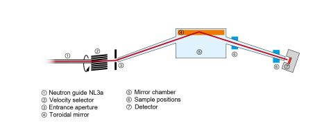
KWS-3 is a very small angle neutron scattering (VSANS) instrument running on the focussing mirror principle. The instrument is designed to bridge the gap between Bonse-Hart and pinhole cameras. Some details of the diffractometer operation are explained in fig. 1 (see gallery): the principle of this instrument is a one-to-one image of an entrance aperture onto a 2D position-sensitive detector by neutron reflection from a double-focussing toroidal mirror.
The instrument’s standard configuration “VSANS” with a 9.3 m sample-to-detector-distance (SDD) and 2 × 2 mm2 entrance aperture (EA) allows performing scattering experiments with a wave vector transfer resolution between 1.0 ×10⁻⁴ and 2.5 ×10⁻³ Å-1.
‘VerySANS’ is a focussing instrument. Unlike at a pinhole SANS instrument, we can not improve the dynamical range (Qmax/Qmin) by moving the detector: the position of the KWS-3 detector is fixed in the right focus as shown in fig. 1. At KWS-3, we extend the dynamical range by ‘replicating’ of the sample positions at different distances between the sample and the detector (fig. 2).
Compact sample environment (SE) (gallery, tab. 1 and fig. 3a): We can install a compact SE (volume below 30 × 30 × 30 cm3) inside two vacuum chambers located at SDD = 9.3 and 1.7 m; as well as those samples can be measured in the close vicinity of the detector (SDD = 0.05 – 0.40 m). In tab. 1, all parameters of configurations of the compact SE are listed. The combination of several configurations, called “USANS”, “VSANS”, “SANS-overlap”, and “SANS”, allows performing scattering experiments with a wave vector transfer resolution between 3.5 ×10⁻⁵ and 3.5 ×10⁻¹ Å-1, covering four decay of the dynamical range.
Bulky sample environment (SE) (gallery, tab. 2 und fig. 3a): In the current instrument configuration, we can install a bulky SE at SDD = 10, 4, 3, and 2 m. At these positions, we can precisely position as well as rotate and tilt a sample together with a bulky/ heavy SE. A sample in a few Tesla magnetic field or under shear in the rheometer can be measured within q-range between 3.5 ×10⁻⁵ and 10-2 Å-1.
The instrument covers the Q range of small angle light scattering instruments. Especially when samples are turbid due to multiple light scattering, VSANS gives access to the structural investigation. Thus, the samples do not need to be diluted. The contrast variation method allows for the highlighting of particular components.
Small angle scattering is used to analyse structures with sizes just above the atomic scale, between 1 and about 100 nm, which cannot be assessed or sufficiently characterised by microscopic techniques. KWS-3 is an important instrument extending the accessible range of scattering angles to very small angles with a superior neutron flux compared to a conventional instrumental set-up with pinhole geometry. Thus, the length scale that can be analysed is extended beyond 10 μm for numerous materials from physics, chemistry, materials science, and life science, such as alloys, diluted chemical solutions, and membrane systems.
- Aggregation in colloidal dispersions
- Self-assembling of polymers
- Hierarchical structures of biominerals
- Hydrogels and aerogels
- Membrane systems
- Rheology and structure/morphology of complex fluids
- Morphology of vortex lattice domains
- Surface and interface structure
- Porous structure & gas storage in rocks
- In-situ study of solid phase transition/crystallisation at high temperature
- Colloid science: mixtures of particles, particles of micron size, silicon macropore arrays
- Materials science: filled polymers, cements, microporous media
- Polymer science: constrained systems, emulsion polymerisation
- Bioscience: aggregations of bio-molecules, protein complexes, crystallisation of proteins
- Multilamellar vesicles
- Neutron polarisation & polarisation analysis
- Anton-Paar fluid rheometer
- Stopped flow mixer with UV-Vis and fluorescence detector
- Different RT-sample holders
- Oil & water thermostats: typical 5 – 150°C
- Electric thermostat: RT – 200°C
- 6/8-positions Peltier sample holder: -10 – 150°C
- Magnet: 2 T, vertical
- Magnet: 3 T, horizontal
- Cryostat with sapphire windows
- High temperature furnace: RT – 2000°C
- Pressure cells: 500 bar, 2000 bar, 5000 bar
- Humidity generator and humidity cells: 0 – 90 RH %
- Linkam Modular Force Stage: tensile testing at various temperatures and humidity
- MgLi velocity selector
- Wavelength spread Δλ/λ = 0.17
- Wavelength range λ = 10.5 – 30 Å (maximal flux at 12.8 Å)
- 0 × 0 – 10 × 10 mm2
- 0.7 × 0.7 mm2 (‘USANS’ modes with VHRD)
- 2.0 × 2.0 mm2 (‘VSANS’ and ‘SANS’ modes with HRD: ‘high resolution’ mode)
- 4.0 × 4.0 mm2 (‘VSANS’ and ‘SANS’ modes with HRD: ‘high intensity’ mode)
- HRD:
- Type: scintillator, 6Li, 1 mm
- Active area: ∅ 9.0 cm
- Pixel size: 0.34 : 0.34 mm2
- Dead-time: 2.9 μs
- Matrix Dim.: 256 × 256
- VHRD:
- Type: scintillator, 6Li, 1 mm
- Active area: 3 × 3 cm2
- Pixel size: 0.12 : 0.12 mm2
- Dead-time: 2.7 μs
- Matrix Dim.: 256 × 256
- 3.5 × 10⁻⁵ – 3.5 × 10⁻¹ Å-1 (compact SE, tab. 1)
- 3.5 × 10⁻³ – 1.2 × 10⁻² Å-1 (bulky SE:, tab. 2)
- Several samples measured at KWS-3 are shown in fig. 3b (see gallery)
- All possible instrument configurations are listed in tab. 1 and 2 (see gallery)
- ‘USANS’, ‘USANS-10m’ and ‘SANS’ configurations:
- Currently only very strongly scattering samples can be measured
- Simulated intensity of ‘VSANS’ and ‘SANS-overlap’ configurations
- 1200 n cm-2 s-1 (in ‘high resolution’ mode with EA = 2 × 2 mm2)
- 4800 n cm-2 s-1 (in ‘high intensity’ mode with EA = 4 × 4 mm2)
- MgLi velocity selector
- Wavelength spread Δλ/λ = 0.17
- Wavelength range λ = 10.5 – 30 Å (maximal flux at 12.8 Å)
- 0 × 0 – 10 × 10 mm2
- 0.7 × 0.7 mm2 (‘USANS’ modes with VHRD)
- 2.0 × 2.0 mm2 (‘VSANS’ and ‘SANS’ modes with HRD: ‘high resolution’ mode) (‘VSANS’ and ‘SANS’ modes with HRD: ‘high intensity’ mode)
- 4.0 × 4.0 mm2 (‘VSANS’ and ‘SANS’ modes with HRD: ‘high intensity’ mode)
- HRD:
- Type: scintillator, 6Li, 1 mm
- Active area: ∅ 9.0 cm
- Pixel size: 0.34 : 0.34 mm2
- Dead-time: 2.9 μs
- Matrix Dim.: 256 × 256
- VHRD:
- Type: scintillator, 6Li, 1 mm
- Active area: 3 × 3 cm2
- Pixel size: 0.12 : 0.12 mm2
- Dead-time: 2.7 μs
- Matrix Dim.: 256 × 256
- 3.5 ×10⁻⁵ – 3.5 ×10⁻¹ Å-1 (compact SE, tab. 1)
- 3.5 ×10⁻³ – 1.2 ×10⁻² Å-1 (bulky SE:, tab. 2)
- Several samples measured at KWS-3 are shown in fig. 3b (see gallery)
- All possible instrument configurations are listed in tab. 1 and 2 (see gallery)
- With the cold source, all instrument configurations are usable.
- Measured flux as a function of the instrument configuration (EA size)
- 3000 n cm-2 s-1 (‘USANS’ mode with EA = 0.7 × 0.7 mm2)
- 24000 n cm-2 s-1 (in ‘high resolution’ mode with EA = 2 × 2 mm2)
- 96000 n cm-2 s-1 (in ‘high intensity’ mode with EA = 4 × 4 mm2)
Instrument scientists
Dr. Vitaliy Pipich
Phone: +49 (0)89 158860-710
E-mail: v.pipich@fz-juelich.de
Dr. Baohu Wu
Phone: +49 (0)89 158860-687
E-mail: ba.wu@fz-juelich.de
KWS-3
Phone: +49 (0)89 158860-513
Operated by

Funding

Publications
Find the latest publications regarding KWS-3 in our publication database iMPULSE:
Citation templates for users
In all publications based on experiments on this instrument, you must provide some acknowledgements. To make your work easier, we have prepared all the necessary templates for you on this page.
Instrument control
Gallery


MLZ is a cooperation between:
 > Technische Universität München
 > Helmholtz-Zentrum Hereon
 > Forschungszentrum Jülich
MLZ is a member of:
 > LENS
 > ERF-AISBL
MLZ on social media:


