MLZ is a cooperation between:
 > Technische Universität München
 > Helmholtz-Zentrum Hereon
 > Forschungszentrum Jülich
MLZ is a member of:
 > LENS
 > ERF-AISBL
MLZ on social media:

MLZ (eng)
Lichtenbergstr.1
85748 Garching
08.06.2021
To the tumor via magnet

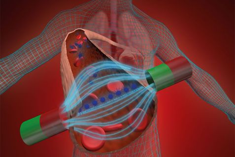
Visual representation of the magnet-induced self-organization of iron oxide nanoparticles (blue) into chains in a blood vessel. © Reiner Müller / FRM II, TUM
Biocompatible iron oxide nanoparticles (IONPs) offer great potential for biomedical applications, both in terms of imaging and therapy. More rapid progress in researching IONPs now looks promising by using a new method combination developed by a team of Jülich researchers using neutrons at the MLZ as a probe.
This makes it possible for the first time to make chains of iron oxide nanoparticles directly visible in dispersions. As a result, it is quicker and easier to determine how parameters such as size, concentration, composition and magnetic fields influence the formation of the chains through self-organization. Up until now, the IONPs were only directly visible when using an electron microscope. For this to work, however, they would need to be deposited to a sample carrier that can influence self-organization.
Use as contrast agent and potential cancer drug
Magnetic nanoparticles can be used in three main biomedical areas: imaging, sensors and therapeutics. As contrast agents in magnetic resonance imaging (MRI) examinations, for example, they are used to make certain tissue structures more visible. For laboratory tests, they can be coupled with antigens to detect antibodies in body fluids, for instance. Various therapeutic approaches are still predominantly in the experimental stage: in tumour tissues, magnetic particles are able to kill cancer cells if they are set rotating using alternating magnetic fields, thus generating heat. If transported strategically to specific sites in the body, they can release active substances precisely where they are needed, again activated by magnetic fields. Both methods can potentially keep side effects to a minimum due to accurate targeting.
Chains can move better in blood vessels

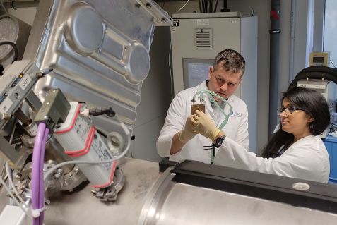
In addition to neutron scattering, researchers also used the Jülich small-angle X-ray diffractometer GALAXI to characterise the iron oxide nanoparticles. GALAXI, pictured here with Nileena Nandakumaran and Dr. Mikhail Feygenson, is operated by the JCNS at a laboratory X-ray source with extremely high brilliance. © Forschungszentrum Jülich
One challenge in medical applications is the strategic transport of the particles to the desired location. Apart from directly injecting the particles at the target site or coating them with organic substances that only bind to certain tissues, another option is to manoeuvre the nanoparticles to the desired position using a magnetic field. Aggregates of the particles are better suited for this than individual nanoparticles because their magnetic moment is greater, which means they can be controlled by smaller magnetic fields. At the same time, thread-like aggregates are advantageous compared to other shapes because the long, thin formations can move better through narrow structures, such as blood vessels.
Finely distributed in a solvent, the nanoparticles can self-assemble into the thread-like chains when conditions are suitable. If there is no spatial boundary acting on the particles, certain parameters such as size, concentration and the composition of the nanoparticles, as well as the strength of the applied magnetic field, determine the self-organization. However, the only method up to now that could directly make the tiny chains visible and track their formation could not function without spatial limitations: For studies involving an electron microscope, samples must be placed on a carrier whose lattice structure provides spatial limitation and is thus able to influence the self-organization of the IONPs.
Neutrons make the chains of nanoparticles visible
Now researchers from Forschungszentrum Jülich, RWTH Aachen University and other German and international research institutions are presenting a new approach that makes the chains and their formation directly visible in the solvent without the need for spatial limitation. To do this, they combined experimental studies using small-angle neutron scattering at the instrument KWS-1 with reverse Monte Carlo simulations. Small-angle scattering enables similarly high-resolution structural analyses to be made as with electron microscopy, but generates data that are not intuitively comprehensible and must be interpreted using elaborate procedures. With the help of simulations, directly recognisable images of the particle chains can now be obtained.
“Reverse Monte Carlo simulation is an established method, but this is the first time we have used it to interpret small-angle scattering data,” reports Nileena Nandakumaran, a PhD student at the Jülich Centre for Neutron Science. Her supervisor, Dr. Mikhail Feygenson, adds: “Visualising IONP aggregates in dispersions in real space offers a novel tool for biomedical researchers. This is because the method enables a comparatively simple, rapid investigation of the behaviour of IONPs in solution in a broad parameter space and an unambiguous extraction of the parameters of the equilibrium structures. It also allows the study of geometrically more complex aggregates of IONPs.”Text: Angela Wenzik / Forschungszentrum Jülich
Original publication:
Unravelling Magnetic Nanochain Formation in Dispersion for In Vivo Applications; Nileena Nandakumaran et al.; Adv. Mater. 2021, 202008683, First published: 07 May 2021, DOI: 10.1002/adma.202008683
Further information:
In addition to scientists from Forschungszentrum Jülich and RWTH Aachen University, researchers from Los Alamos and Sandia National Laboratories, USA, the German Electron Synchrotron DESY and the Paul Scherrer Institute, Switzerland, were also involved in the research work.
Contact:
Nileena Nandakumaran
Jülich Centre for Neutron Science (JCNS)
Forschungszentrum Jülich
Phone: 02461 61-4037
E-Mail: n.nandakumaran@fz-juelich.de
Press contact:
Angela Wenzik
Wissenschaftsjournalistin
Forschungszentrum Jülich
Phone: 02461 61-6048
E-Mail: a.wenzik@fz-juelich.de
MLZ is a cooperation between:
 > Technische Universität München
 > Helmholtz-Zentrum Hereon
 > Forschungszentrum Jülich
MLZ is a member of:
 > LENS
 > ERF-AISBL
MLZ on social media:


