MLZ is a cooperation between:
MLZ is a member of:
 > ERF-AISBL
MLZ on social media:


MLZ (eng)
Lichtenbergstr.1
85748 Garching
12.06.2023
From Orange to Red: How bacteria protect themselves from sunlight
Cyanobacteria have a built-in sunscreen in their cells. Using neutrons at MLZ, a German-Estonian research team has now directly observed the orange carotenoid protein in action. Surprisingly, they found that a previously much-studied mutant of the protein behaves significantly differently than the original.

Dr. Wiebke Lohstroh at the time-of-fligth-spectrometer TOFTOF, inserting a sample tube © Andreas Heddergott, FRM II / TUM
Proteins are one of the most important building blocks in living organisms. In bacteria, they protect sensitive cell components, among other things. Proteins are made up of long-chain bonds of amino acids that fold into complex three-dimensional arrangements. This structure gives them the ability to carry out transport processes, metabolism, and communication between individual cell components. Some proteins change their shape in response to certain external stimuli to carry out their task. This is the case with the orange carotenoid protein (OCP) found in cyanobacteria.
Red instead of orange when exposed to too much sun
The unicellular organisms, which are often found in freshwater, the sea, or the ground and are incorrectly called blue-green algae, carry out photosynthesis just like plants. If the sunlight becomes too strong, it will damage the reaction centers in the bacteria. The biological trick to prevent this lies in the OCP. If the protein perceives excess light, it changes from its inactive orange form (OCP-O) to the active red form (OCP-R) and attaches itself to the light-processing cell components. The active form itself absorbs light and converts it into heat – the load on the photosynthetic machinery decreases.
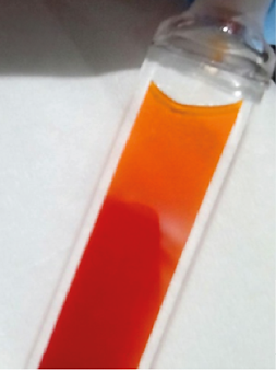
Phase separation between orange (inactive) and red (active) proteins in a glass vial. Activation occurs upon illumination.
Getting to the heart of proteins with neutrons
The focus of the investigation was on the dynamic processes inside the protein and their role in the transition to the active state, which has been little studied to date. A specific wavelength is selected from the neutrons at FRM II on the time-of-flight spectrometer TOFTOF. The neutrons strike the sample, changing its wavelength. From this change, the researchers conclude the movement of the proteins.
“OCP is very well characterized and allows us to perform successive measurements with and without light on the same sample,” says Dr. Wiebke Lohstroh, a scientist at the TOFTOF time-of-flight spectrometer at the Heinz Maier-Leibniz Research Neutron Source (FRM II).
While the atomic structure of the inactive protein configuration OCP-O in solution is known, there is little knowledge about the corresponding structure of the active OCP-R, although it is precisely the structure that determines the dynamic behavior. The mutant protein OCP-W288A, on the other hand, has been extensively characterized and is so far considered a good model system for the solubilized OCP-R. The researchers thus integrated the mutant into their experiments as an object of comparison. To stimulate and subsequently measure the protein’s change of state, Dr. Maksym Golub and Prof. Dr. Jörg Pieper from the University of Tartu, Estonia, teamed up with Lohstroh. Together, they supplemented the TOFTOF with an optical setup that can irradiate a sample cell with light.
Small movements – big effect
For the first time, Golub and Pieper’s team was able to directly observe the lower stability of the active state OCP-R in an in situ experiment. Thus, the average time that the active protein dwells in each state is noticeably shorter than that of the inactive variant. This can be explained by an “opening” of the protein during activation. The surface area increases, allowing more water molecules to collide with it. Contrary to previous assumptions, the mobility of the protein mutant OCP-W288A proved to be significantly greater once again, which greatly limits its comparability with OCP-R.
This again demonstrates the ability of scientific research to constantly evolve and critically question previously made assumptions. The comparative values obtained here may prove valuable for follow-up studies that seek to simulate the molecular dynamics of these proteins. “Without this experiment, we would not know how much more unstable the mutation makes the OCP,” Lohstroh explains. “This knowledge will now help us understand proteins even better in the future.”
Original publication:
M. Golub, M. Moldenhauer, F.-J. Schmitt, W. Lohstroh, T. Friedrich, and J. Pieper
Light-Induced Conformational Flexibility of the Orange Carotenoid Protein Studied by Quasielastic Neutron Scattering with In Situ Illumination
J. Phys. Chem. Lett. 2023, 14, 295−301
10.1021/acs.jpclett.2c03198
More information:
The project received financial support from the Estonian Research Council (Grants PRG 539 and SLOKT 12026 T) and the German Research Foundation (DFG, Grants FR 1276/5-1 and FR 1276/6-1 to T.F.).
In addition to the Technical University of Munich and the University of Tartu, researchers from the Martin Luther University of Halle and the Technical University of Berlin were also involved.
Contact:
Prof Dr Jörg Pieper
Institute of Physics, University of Tartu, Estonia
Phone: +(372) 737 4627
E-Mail: pieper@ut.ee
Related News


MLZ is a cooperation between:
MLZ is a member of:
> ERF-AISBL
MLZ on social media: